
| IMGT Web resources |
|
| Here you are: IMGT Web resources > IMGT Education > Tutorials > Immunoglobulins and B cells |
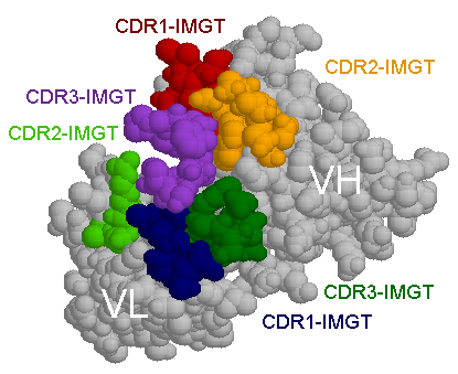
The VL and VH domains have been moved to show the CDR loops.
Reference:
| [1] | Padlan E.A., Mol.immunol. 31, 169-217 (1994) |
| [2] | Padlan E.A., Adv.Protein.Chem 49, 57-133 (1996) |
| [3] | Webster D.M. et al., Curr.Opin.Struct.Biol. 4, 123-129 (1994) |
| [4] | Wu T.T et al., Proteins: Struct.Funct.Genet. 16, 1-7 (1993) |
The corresponding PDB (Protein Data Bank) format file is from Mike's immunoglobulin structure function site (http://www.umass.edu/microbio/rasmol/padlan.htm).
Created: 28/06/2001
Last updated: Friday, 06-Feb-2026 15:23:28 CET
Author: Marie-Paule Lefranc
Marie-Paule.Lefranc@igh.cnrs.fr
Editors: Elodie Foulquier, Chantal Ginestoux
IMGT Home page |
IMGT Repertoire (IG and TR) |
IMGT Repertoire (MH) |
IMGT Repertoire (RPI) |
IMGT Index |
IMGT Scientific chart |
IMGT Education |
IMGT Latest news ![]()


© Copyright 1995-2026 IMGT®, the international ImMunoGeneTics information system® | Terms of use | About us | Contact us | Citing IMGT