
| IMGT Web resources |
|
| Here you are: IMGT Web resources > IMGT Education > Tutorials > Immunoglobulins and B cells |
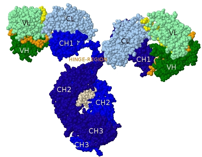
The first crystal 3D structure of a complete human immunoglobulin is that of b12, a human IgG1, anti-gp120 [HIV-1], determined in 2001 (PDB code: 1hzh in IMGT/3Dstructure-DB).
Archive: Spacefill 3D representation of an IgG model
Created: 28/06/2001
Last updated: Friday, 06-Feb-2026 15:23:27 CET
Author: Marie-Paule Lefranc
Marie-Paule.Lefranc@igh.cnrs.fr
Editors: Elodie Foulquier, Chantal Ginestoux
IMGT Home page |
IMGT Repertoire (IG and TR) |
IMGT Repertoire (MH) |
IMGT Repertoire (RPI) |
IMGT Index |
IMGT Scientific chart |
IMGT Education |
IMGT Latest news ![]()


© Copyright 1995-2026 IMGT®, the international ImMunoGeneTics information system® | Terms of use | About us | Contact us | Citing IMGT