Half-IG exchange
Human IgG4 form bispecific antibodies through a dynamic half-IG exchange process, that occurs naturally in vivo. In this process, half-IG of an IgG4 is exchanged with half-IG (i.e. one heavy H-GAMMA-4 and one light chain) of another IgG4 [1].
This bispecific antibody is monovalent with respect to each specificity and was proposed to contribute to the anti-inflammatory properties of IgG4 [2].
This process is also called 'Fab-arm exchange' in the literature but this expression is inaccurate since the hinge, CH2 and CH3 are also exchanged. Half-IG exchange has been used in antibody engineering to generate stable bispecific IgG1.
Half-IG exchange in human IgG4
Heavy (H) H-GAMMA-4 chains of human immunoglobulin IgG4 are covalently linked by two disulfide bridges located in their hinge region (involving the cysteines (Cys) C8 and C11).
However, it has been shown that the two inter-H disulfide bonds are in equilibrium with intra-chain disulfide bonds [3].
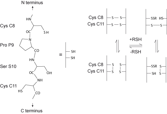
Figure 1. Schematic representation of the equilibrium between the different IgG4 disulfide bond isoforms with RSH representing a sulfhydryl reducing agent like dithiothreitol (DTT), reduced glutathione (GSH), β-mercaptoethanol (BME) (modified from Rispens et al. [4]).
The part of the hinge amino acid sequence with the cysteines Cys C8 and C11 is represented on the left handside (for the IMGT numbering see the Correspondence between C numberings).
Two amino acids have been shown to play a key role in this half-IG exchange: the serine (Ser) S10 of the hinge region (instead of proline (Pro) P10 in IgG1) and the arginine (Arg) R88 (instead of lysine (Lys) K88 in IgG1) at the interface of the CH3 domain.
S10 of the H-GAMMA-4 hinge region, introduces flexibility and is thought to make the inter-H disulfide bridges more easily reduced [5].
R88 in the E-STRAND of CH3 reduces the strength of the CH3-CH3 interaction in IgG4, and allows this subclass to engage half-IG exchange [6], [7].
The 3D structure of the human IgG b12 antibody is shown here with the hinge region (containing P10) and the CH3 domains (containing K88) labeled in red (1hzh in IMGT/3Dstructure-DB).
The half-IG exchange event occurs also for therapeutic IgG4 antibodies.
Indeed, natalizumab has been shown to exchange half-IG with endogenous human IgG4 in natalizumab-treated patients.
To decrease the half-IG exchange of therapeutic antibodies, a S10>P change is introduced in the hinge region in order to obtain hinge-stabilized IgG4 antibodies (e.g. gemtuzumab), which make them less succeptible to half-IG exchange in vivo [8].
Strategy for the generation of bispecific IgG1 by controlled half-IG exchange
mAb1 and mAb2 represent two antibodies with different specificities.
VH-1 and VL-1 represent the VH and VL domains of mAb1, whereas VH-2 and VL-2 represent the VH and VL domains of mAb2.
Amino acid changes at position 88 and 85.1 at the interface between the CH3 domains of the two H-GAMMA-1 are made (K88>R in mAb1 and F85.1>L in mAb2). The AA changes decrease the interaction strength between the H-GAMMA-1 of both homodimers mAb but are expected to increase the interaction strength in a heterodimer [9].
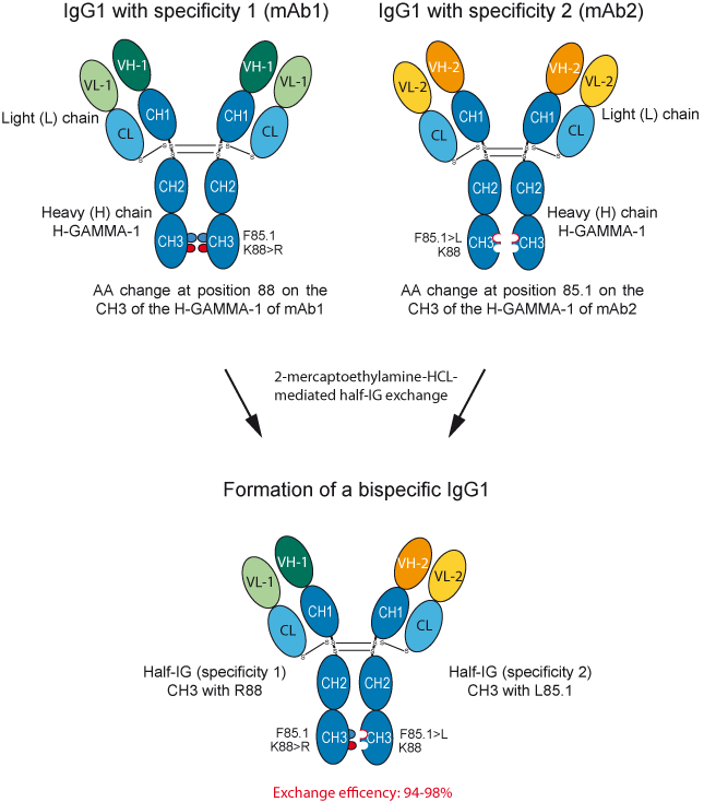
Strategy for obtaining bispecific IgG1 using the controlled half-IG exchange.
The two engineered parental antibodies, each containing a single amino acid change in CH3 are first expressed separately, and then recombined using a reducing agent which mediates half-IG exchange. The efficiency of half-IG exchange ranges between 94 and 98% [9].
AA positions in IMGT Collier de Perles
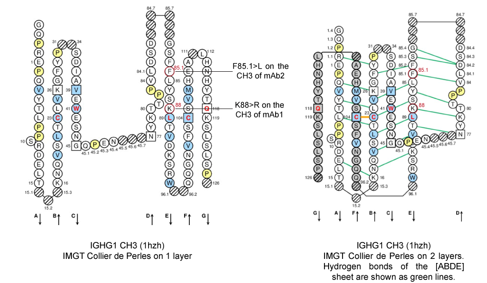
IMGT Collier de Perles from Homo sapiens IGHG1 CH3 (1hzh in IMGT/3Dstructure-DB).
K88 and F85.1, both in E-STRAND of the CH3 of H-GAMMA-1 of an IgG1 are shown in the IMGT Collier de Perles, in red circles.
The E-STRAND belongs to the [ABDE] sheet at the interface between the two CH3 (IMGT Collier de Perles on 2 layers).
Molecular interactions at the interface between the two CH3 domains
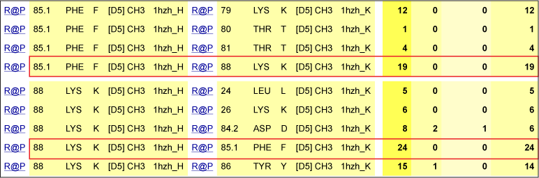
In the interface between the CH3, the lysine (Lys) K88 in E-STRAND is within close proximity with the phenylalanine (Phe) F85.1 in E-STRAND on the partner CH3.
K88 makes numerous van der Waals contacts with F85.1 (red box). However, K88 also makes numerous contacts with L24, K26, D84.2 and Y86 on the other domain.
Contacts between K88 and F85.1 of the CH3 domain of H-GAMMA-1 of an IgG1 (1hzh in IMGT/3Dstructure-DB).
AA positions in the 3D structure
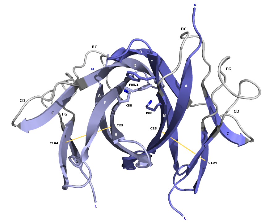
3D structure of the CH3 dimer interface from the human IGHG1 (1hzh in IMGT/3Dstructure-DB).
Amino acids K88 and F85.1 that are mutated (K88>R in mAb1, F85.1>L in mAb2) in the half-IG exchange are represented with their side chains. Intrachain disulfide bridges between C23 (1ST-CYS) in B-STRAND and C104 (2ND-CYS) in F-STRAND are shown in yellow [9].
- Amino acid positions involved in ADCC, ADCP, CDC, half-life and half-IG exchange (IMGT Education> Tutorials)
| [1] | Schuurman, J. et al. Immnunology, 97(4):693-8 (1999) PMID: 10457225 |
| [2] | van der Neut Kolfschoten, M. et al. Science, 317(5844):1554-7 (2007) PMID: 17872445 |
| [3] | Schuurman, J. et al. Mol Immunol. 38(1):1-8.(2001) PMID: 11483205 |
| [4] | Rispens, T. et al. J Am Chem Soc. 133(26):10302-11 (2011) PMID: 21627172 |
| [5] | Bloom, J. W. et al. Protein Sci. 6(2):407-15 (1997) PMID: 9041643 |
| [6] | Labrijn, A. F. et al. J Immunol. 187(6):3238-46 (2011) PMID: 21841137 |
| [7] | Davies, A.M. et al. Mol Immunol. 54(1):1-7 (2013) PMID: 23164605 |
| [8] | Labrijn, A. F. et al. Nat Biotechnol. 27(8):767-71 (2009) PMID: 19620983 |
| [9] | Labrijn, A. F. et al. Proc Natl Acad Sci U S A. 110(13):5145-50 (2013) PMID: 23479652 |
IMGT Home page |
IMGT Repertoire (IG and TR) |
IMGT Repertoire (MH) |
IMGT Repertoire (RPI) |
IMGT Index |
IMGT Scientific chart |
IMGT Education |
IMGT Latest news ![]()


© Copyright 1995-2026 IMGT®, the international ImMunoGeneTics information system® | Terms of use | About us | Contact us | Citing IMGT