3D representation: Human
V-DOMAINs from TcR ab A6 (PDB: 1ao7 )
IMGT/3Dstructure-DB card: 1ao7
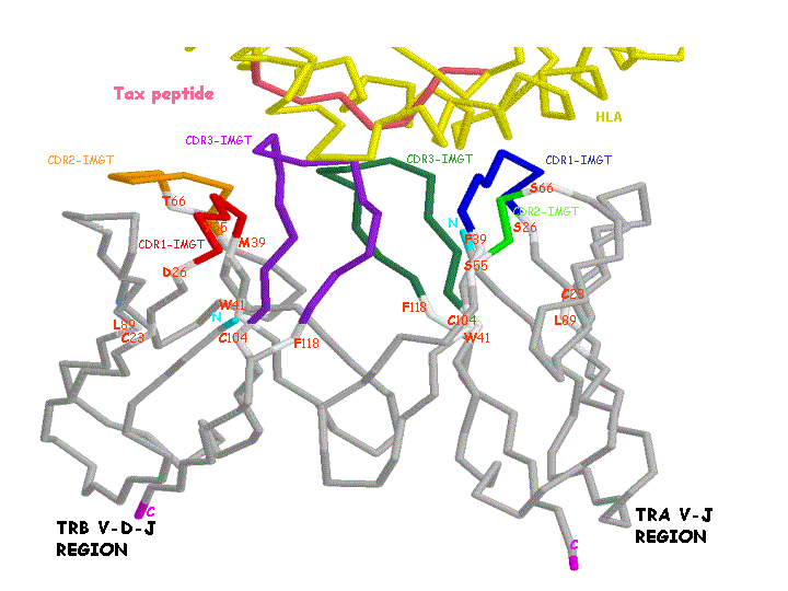
Side view (backbone model)
View from above the CDRs (backbone model)
Collier de perles: Human TRA V-J REGION
Collier de perles: Human TRB V-D-J REGION
Complex TcRabA6-Tax/HLA-A*0201 (backbone model)

Legend:
- CDR-IMGT regions are colored according to the IMGT color menu:
TRA V-J-REGION: CDR1-IMGT (blue), CDR2-IMGT (green), CDR3-IMGT (greenblue)
TRB V-D-J-REGION: CDR1-IMGT (red), CDR2-IMGT (orange), CDR3-IMGT (purple)
The N (cyan) and C (magenta) ends of each region are also colored.
- The conserved C 23, W 41 and L 89, and the FR-IMGT amino acids limiting the CDR-IMGT regions (positions 26 and 39 for the CDR1-IMGT, 55 and 66 for the CDR2-IMGT, 104 and 118 for the CDR3-IMGT) are shown in white.
- This image has been generated with the program RASMOL version 2.6 [1] from the PDB file 1a07 [2], containing the atomic coordinates of the complex between human TcR ab A6, viral peptide Tax and human MHC molecule HLA-A*0201. Only the human TcR ab A6 V-DOMAINs, the peptide Tax (in red) and parts of the HLA-A alpha1 and alpha2 helices (in yellow) are displayed here.
References:
[1] Sayle, R. and Milner-White, E.J. TIBS, 20, 374 (1995).
[2] Garboczi, D.N. et al., Nature, 384, 134 (1996).
Created: 11/06/2001
Authors: Manuel Ruiz and Marie-Paule Lefranc
Editors: Elodie Foulquier and Marie-Paule Lefranc

