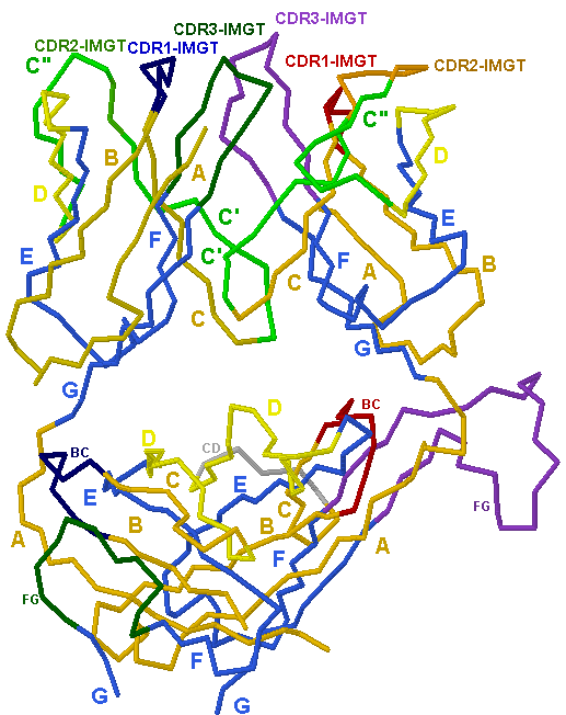
TR αβ B7 (PDB: 1bd2)
IMGT/3Dstructure-DB card: 1bd2
Side view (backbone model)
| TRA V-DOMAIN V-ALPHA TRA V-J-REGION |

|
TRB V-DOMAIN V-BETA TRB V-D-J-REGION |
| TRAC C-DOMAIN C-ALPHA |
TRBC2 C-DOMAIN C-BETA |
This figure shows the two associated V-DOMAINs and C-DOMAINs of an alpha-beta T cell receptor (alpha chain (PDB: 1bd2_D), delta chain (PDB: 1bd2_E).
The two V-DOMAINs, V-ALPHA and V-DELTA, correspond to the TRA V-J-REGION and TRB V-D-J-REGION, respectively.
The two C-DOMAINs, C-ALPHA and C-BETA2, are part of the TRA C-REGION and TRB C-REGION, respectively.
Only the extracellular domains are displayed. The carbohydrates are not shown.
In the PDB file, the sequences include 10 and 17 amino acids of the CONNECTING-REGION of the alpha and delta chains, respectively,
however, the 3D structures only include amino acids up to positions 203 and 247, for the alpha and beta C-DOMAINs, respectively,
according to the IMGT unique numbering for C-DOMAIN.
The TRAV-J rearrangement is the following: TRAV-TRAJP (Human TRAV cDNAs).
The TRBV-D-J rearrangement is the following: TRBV6-5-TRBD2-TRBJ2-7 (Human TRB cDNAs).
- V-DOMAIN CDR-IMGT regions and C-DOMAIN loops are colored according to the IMGT color menu:
- V-DOMAIN and C-DOMAIN strands are colored according to the IMGT color menu for strand representation: A, B and C strands (orange), D strand (yellow), E, F and G strands (blue).
- V-DOMAIN C' and C'' strands (green)
- C-DOMAIN transversal CD strand (grey)
This image has been generated with the program RASMOL version 2.6 [1] from the PDB file 1bd2 [2], containing the atomic coordinates of the complexe of a human T cell receptor B7.
References:
[1] Sayle, R. and Milner-White, E.J. TIBS, 20, 374 (1995).
[2] Ding, Y.H. et al., Immunity, 8, 403-411 (1998).


